
You have no items in your shopping cart.
All Departments
- Home Home
-
SpO2 Sensors
SpO2 Sensors

-
ECG Cables
ECG Cables

-
EKG Cables
EKG Cables

-
NIBP Cuffs
NIBP Cuffs

- EtCO2 Sensors EtCO2 Sensors
- IBP Cables IBP Cables
- Temperature Probes Temperature Probes
- Fetal Probes Fetal Probes
- Patient Monitors Patient Monitors
- Medical Accessories Medical Accessories
- Veterinary Accessories Veterinary Accessories
- ESU Accessories ESU Accessories
- oxygen sensor oxygen sensor
Veterinary ECG Leads, Blood Pressures and SP02 Sensor Placement
# Veterinary ECG Lead Placement
# Veterinary Blood Pressure Cuff Placement
# Veterinary SPO2 Sensor Placement
1.Veterinary ECG Lead Placement
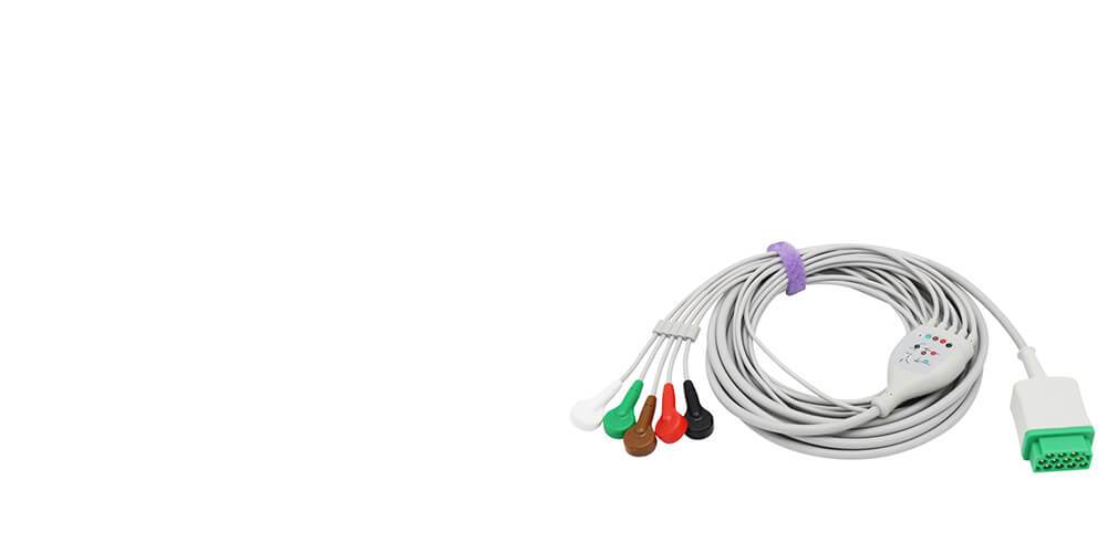
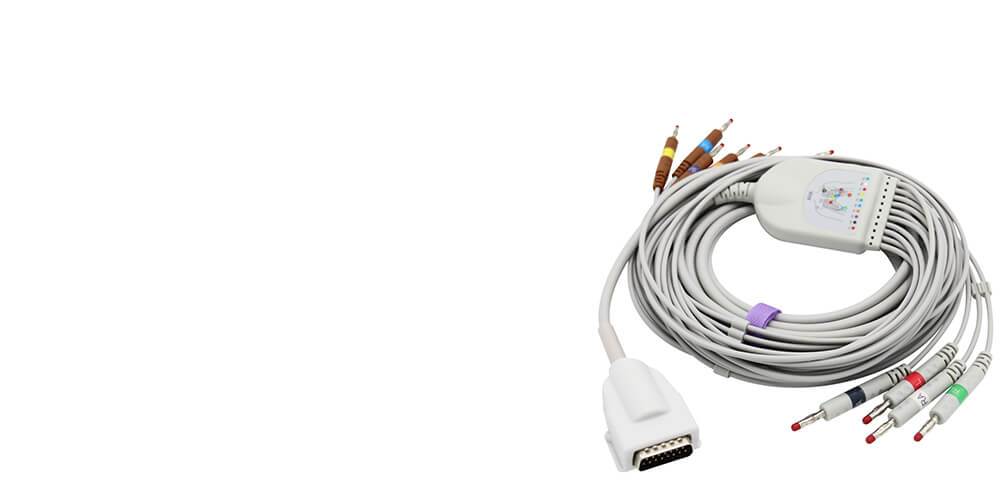
An electrocardiogram (ECG) can be a critical tool in diagnosing and managing heart disease in dogs and cats. However, interpreting an ECG can be challenging even with the best recording. So here is 3 Lead Placement
Lead 1: LA - RA: Left foreleg (left arm) electrode (+) placed just below the point of the elbow on the back of the left forearm - right foreleg (right arm) electrode (-) placed just below the point of the elbow on the back of the right forearm.
Lead II: LL - LA: Left hindleg (left leg) electrode (+) placed on the loose skin at the left stifle in the region of the patella - left foreleg (left arm) electrode (-) placed just below the point of the elbow on the back of the left forearm.
Lead III: LL - RA: Left hindleg (left leg) electrode (+) placed on the loose skin at the left stifle in the region of the patella - right foreleg (right arm) electrode (-) placed just below the point of the elbow on the back of the right forearm.
For example: ECG Placement on Dog
2. Veterinary Blood Pressure Cuff Placement
Choose a cuff whose width is 40%-50% of limb circumference of the limb to which you will attach it, and round up to the next sized cuff if in between. The inflatable part of the cuff (bladder) should be long enough to encircle 50%-80% of limb.
For example: Blood Pressure Cuff Placement on Dog

The size of the cuff is important and should measure 40% of the appendage circumference in dogs and 30% in cats. Using a cuff that is too large will typically give falsely low blood pressure readings, while the opposite is true for a cuff that is too small.
The top of the cuff is 23cm below the right atrium.
23cm X 0.8mmHg = 18.4mmHg
Corrected value:
160mmHg- 18.4mmHg = 141.6mmHg
If oscillometric machines are used repeat steps 1-3 and record readings. Use corrected value equation if patient is standing or sitting.
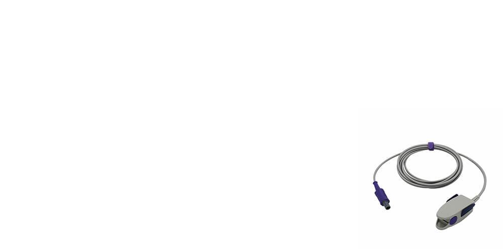
3. Veterinary SPO2 Sensor Placement
A pulse oximeter has a probe which is placed on the patient. The common locations include hairless, minimally-pigmented areas of the body including the lip, pinnae, prepuce, vulva, and interdigital space. Essentially, the probe can be placed on any area with a pulsating arteriolar bed.
Transillumination pulse oximetry of the tongue has been documented as a viable method of monitoring, but not everyone has the equipment available to place a transilluminating pulse oximeter on the tongue. A reflectance pulse oximeter sensor has the light source and the light detector in a side-by-side configuration rather than in opposition.
Veterinary ECG Leads, Blood Pressures and SP02 Sensor Placement
# Veterinary ECG Lead Placement
# Veterinary Blood Pressure Cuff Placement
# Veterinary SPO2 Sensor Placement
1.Veterinary ECG Lead Placement
An electrocardiogram (ECG) can be a critical tool in diagnosing and managing heart disease in dogs and cats. However, interpreting an ECG can be challenging even with the best recording. So here is 3 Lead Placement
Lead 1: LA - RA: Left foreleg (left arm) electrode (+) placed just below the point of the elbow on the back of the left forearm - right foreleg (right arm) electrode (-) placed just below the point of the elbow on the back of the right forearm.
Lead II: LL - LA: Left hindleg (left leg) electrode (+) placed on the loose skin at the left stifle in the region of the patella - left foreleg (left arm) electrode (-) placed just below the point of the elbow on the back of the left forearm.
Lead III: LL - RA: Left hindleg (left leg) electrode (+) placed on the loose skin at the left stifle in the region of the patella - right foreleg (right arm) electrode (-) placed just below the point of the elbow on the back of the right forearm.
For example: ECG Placement on Dog
2. Veterinary Blood Pressure Cuff Placement
Choose a cuff whose width is 40%-50% of limb circumference of the limb to which you will attach it, and round up to the next sized cuff if in between. The inflatable part of the cuff (bladder) should be long enough to encircle 50%-80% of limb.
For example: Blood Pressure Cuff Placement on Dog
The size of the cuff is important and should measure 40% of the appendage circumference in dogs and 30% in cats. Using a cuff that is too large will typically give falsely low blood pressure readings, while the opposite is true for a cuff that is too small.
The top of the cuff is 23cm below the right atrium.
23cm X 0.8mmHg = 18.4mmHg
Corrected value:
160mmHg- 18.4mmHg = 141.6mmHg
If oscillometric machines are used repeat steps 1-3 and record readings. Use corrected value equation if patient is standing or sitting.
3. Veterinary SPO2 Sensor Placement
A pulse oximeter has a probe which is placed on the patient. The common locations include hairless, minimally-pigmented areas of the body including the lip, pinnae, prepuce, vulva, and interdigital space. Essentially, the probe can be placed on any area with a pulsating arteriolar bed.
Transillumination pulse oximetry of the tongue has been documented as a viable method of monitoring, but not everyone has the equipment available to place a transilluminating pulse oximeter on the tongue. A reflectance pulse oximeter sensor has the light source and the light detector in a side-by-side configuration rather than in opposition.


← Older Post Newer Post →